
The practice of minimally invasive dentistry is a relatively recent change in approach for dental professionals, with a significant amount of positive evidence becoming available on the subject, especially in the last ten years. 1 Novel techniques and materials are fast being developed for dentists to keep up with the demands of a patient population who are increasingly concerned about the appearance of their teeth. With a rise in social media many patients wish to not only preserve their dentition but enhance it. As a young dentist, it's not difficult to see that our current population's expectations of dental treatment are vastly different to what our predecessors who graduated 20-30 years ago faced. With this in mind, it is vital that we as dental professionals stay up to date with new dental innovations.
The ICON (Infiltration CONcept) was designed as a minimally invasive resin infiltration system for treating incipient caries in patients of all ages. 2 The low viscosity unfilled resin, developed by the company DMG (Germany) camouflages white spots by means of optical manipulation, and no tooth tissue removal is strictly necessary.
The clear resin flows into the demineralised enamel, and has similar optical properties (similar refractive index) to the enamel, therefore reflecting light to match the the tooth's natural shade. 3 , 4 , 5
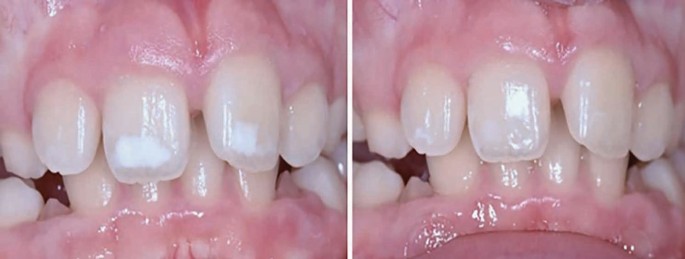
White spot lesions are white opacities seen on teeth after the subsurface layer of enamel on a tooth becomes demineralised, often due to poor oral hygiene and plaque, bacteria and acid accumulation on teeth. The decalcified inner enamel remains trapped underneath remineralised surface enamel. The inner demineralised enamel scatters the light due to its irregular microstructure and the result is an opaque white appearance of the tooth.
White spots can also occur on the teeth due to:
Working in a dental hospital, white spot lesions on anterior teeth are a common presentation that I see on the paediatric department. However, patients will usually present to their primary care practitioner first. Patients of any age can attend your surgery wishing to hear the options for treatment of their white spots, and it is important to be able to distinguish the cause of the white spot, in order to provide the best treatment outcome. If uncertain, a referral for specialist opinion is recommended.
*These treatments are not suitable for children under age of 18 years of age, due to the developing dentition, and the EU regulations for dental bleaching.
A common misconception by dental professionals and patients is that some white spots may be successfully treated with bleaching alone. However, although the overall colour of the tooth will improve, the white spot will remain unchanged and can sometimes even look worse compared to the whiter natural tooth.
A substantial thickness of composite may be required to fully mask a larger white lesion, which can leave the tooth looking bulky. Furthermore, composite and porcelain veneers may require tooth surface preparation which is irreversibly destructive to the tooth.
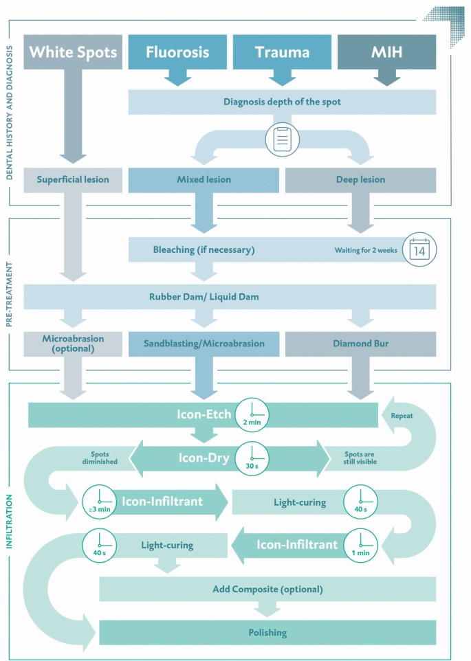
The resin is applied as part of three-part technique. 2 The use of rubber dam is essential to protect the soft tissues. Pre and post-operative photographs are important as part of your record keeping process.
The flow chart in Figure 1, produced by DMG Dental, outlines the process for ICON.
ICON has shown in cases that aesthetic results can be delivered on white spots in a minimally invasive way. 3 , 4 , 5 , 6 This technique should always be used in conjunction with an intensive prevention plan including oral hygiene and diet advice, fluoride varnish placement and regular dental review. Although long term clinical data on this product are limited in the literature, use of this technique is a step forward in allowing patients of all ages to access aesthetic treatment of their white spots, which can significantly impact on their confidence and wellbeing. See Table 1.
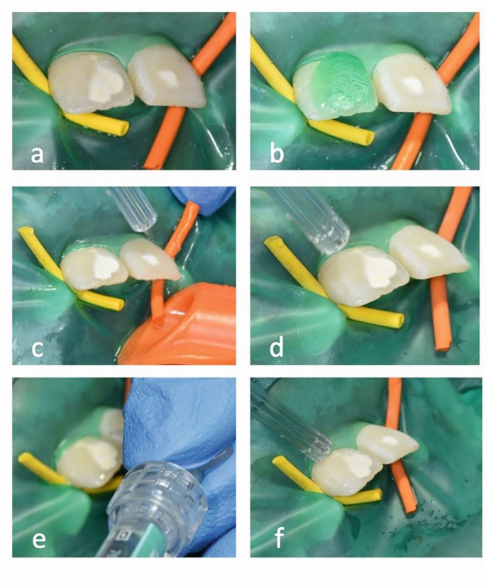
With special thanks to Professor Helen Rodd, Professor/Honorary Consultant in Paediatric Dentistry, the University of Sheffield, and Dr Noren Hasmun, who kindly provided permission for use of clinical photos from her PhD research conducted at the University of Sheffield, UK.
BDS (Manchester), MFDS (Royal College of Physicians and Surgeons of Glasgow). Dental Core Trainee 2 in Paediatric Dentistry at Charles Clifford Dental Hospital, Sheffield, UK
Would you like to submit an original article to BDJ Team for consideration for publication? Just visit our website and click 'submit', or you can email the editor for more information: Kate Quinlan, k.quinlan@nature.com.